Nuclear Medicine use radiopharmaceuticals, which are small amounts of radioactive chemicals, sometimes referred to as radionuclides or radioisotopes. These enable us to see the structure of organs and how they are working.
Within Nuclear Medicine there are three types of services provided:
Most patients who visit us are imaging outpatients. You are usually given a small arm injection of a radiopharmaceutical, no more painful than typical blood test. It is very rare for anyone to experience side-effects.
Children are given anaesthetic cream before they have the injection. They will have a parent or carer with them and there are books, toys and someone to chat with.
You may be asked to sit or lie still on a couch while the camera takes images. You will not be completely enclosed in the camera; it does not make much noise and the gamma camera will move close to you but will never touch you.
The radiopharmaceutical given to you is chosen to target a specific part of the body. We can use this technique to look at a wide variety of organs and diseases. The exact area depends on what the doctor wants to look for.
The camera can see where the radiopharmaceutical has gone and creates a picture of the distribution of radiation in the body. If there is more radiation in one area, this will show up as a darker area in the final image. This can be used to see how an organ functions, for example watching the radiopharmaceutical move through the kidneys.
The most common of these tests is Glomerular Filtration Rate, which is used to see how well the kidneys are functioning. A small amount of radiopharmaceutical is given to you as an injection. Blood samples are then collected.
The amount of radiation in the samples is counted and the results are put into a graph. The graphs are then used to determine whether the samples are within normal limits.
Nuclear medicine may also be used for therapy. Larger amounts of radiopharmaceuticals are given to you and are designed to go to the diseased areas in the body. The radiopharmaceuticals used are different from those used for diagnosis as the radiation kills the diseased tissue. The therapy radioisotopes usually come in small capsules which you swallow.
For some therapies a short stay in hospital is required. However, more than 95% of our therapies are done on an outpatient basis.
What about the radiation dose?
We are all exposed to radiation from natural sources each year. The dose from many of our scans is small, around the average amount we are exposed to in a year. Most of it passes straight through you.
I’ve already had some tests that use radiation, and now I’m having this one. Will the radiation cause me any harm?
We are all surrounded by radiation everyday. The amount of radiation used in this test is very small and very unlikely to cause you any harm. Your doctor thinks that the results of this test are more important than the small risk of radiation.
I’m going abroad on holiday a week after my test, is this OK?
Depending on which test you are having there will be very small amounts of radiation in your body up to two months after the test. Airports and ferry ports have very sensitive detectors that can pick up this radiation. This does not mean that we have to cancel your appointment. Please let the department know if you are planning on going abroad within two weeks of your test
I have a young puppy at home, will the radiation affect him?
Because your puppy is still growing, some of our tests may mean that it would be best for you to avoid prolonged close contact with him until the morning after the test. Our staff will give you advice on how long you should take these precautions for.
I’m babysitting my two grandchildren the day after my test, will this be alright?
For most tests, almost all of the radiation will have gone from your body by the morning after your test is completed. We will advise you on contact restrictions for the next day, including activities such as babysitting the day after the test.
Will the radiation make me feel or look any different?
The radiation is only used to enable us to ‘see’ inside your body using our cameras. The radiation by itself won’t make you look or feel any different.
Our services
Nuclear medicine procedures must be justified, the benefit of having the investigation will far outweigh the risk of the radiation dose. Nuclear medicine is a branch of medicine and medical imaging that uses the radioactive or nuclear properties of certain substances.
There are many different staff groups involved in nuclear medicine, all working as part of a multidisciplinary team. These include the following groups who carry out a range of tasks listed below:
Administrative Staff
When visiting the nuclear medicine department, the first people to greet you will likely be one of our friendly admin team. Their other duties include:
- Attending patients whilst answering all patient and colleague queries
- Managing all of the appointments within the Nuclear Medicine Service and PET/CT service, incorporating the clerical and administrative aspects
Clinical Staff
The staff members who mainly provide care for the patients who visit our department are:
- Clinical Technologists
- Nursing Staff
- Assistant Technical Officers
Individual roles within this staff group will vary, but many tasks will include:
- Administering radiopharmaceuticals to patients
- Dispensing radiopharmaceuticals to be administered to the patient
- Analysing non-imaging investigations in the laboratory
- Processing the images obtained
- Regularly undertaking quality assurance checks on the gamma cameras
- Delivering therapy treatments
- Carrying out patient stressing for cardiac procedures
- Ensuring infection control measures are in place
- Acting as patient advocates
Physics Staff
Physics staff primarily to provide scientific support to the department – this can include:
- Participating in research and development in nuclear medicine
- Giving radiation protection advice to staff, patients and the public
- Processing and reporting complex investigations
- Regularly checking equipment
- Clinical duties such as administering therapeutic agents and performing dosimetry measurements and calculations
Radiologists
- Authorise patient referrals
- Administer radioactive substances (IRMER licence holders)
- Review and report on the images acquired from scans
Radiopharmacy Staff
- Prepare radiopharmaceuticals for patients
- Quality control test the radiopharmaceutical prepared
- Prepare radiopharmaceuticals for external companies
Other staff
There are also a number of other groups of staff that contribute to the nuclear medicine service for example: oncologists, haematologists, service managers, porters, and the ambulance service.
We provide the following services:
- Diagnostic imaging, non-imaging and molecular radiotherapy procedures
- Quality control of equipment, including the gamma cameras used for imaging patients
- Manufacture of radiopharmaceuticals in the purpose-built radiopharmacy
- Radiation protection advice relating to the transport, storage and handling of radioactive materials
- Radiation protection advice for staff, patients and the public following a nuclear medicine investigation
- Advice on compliance with current legislation relating to the administration of radioactive sources to patients
- Application and monitoring of IRMER licences
- Research and development
Each Nuclear Medicine referral form must be filled in correctly and must fulfil all of our requirements before we can accept it. Each referral is reviewed and then authorised by a member of our team in accordance with ARSAC protocols and following IRMER 2017 guidelines.
Therefore, please ensure all referrals contain:
- The patient’s full name, address and date of birth
- The doctor’s ‘wet’ signature and a clearly legible name and date
- The doctor’s contact details
- The name of the patient’s consultant if available
- The test which is being requested
- Clinical history of why the test has been requested
- The required outcome of the test, ie. question to be answered by the test
- Any additional information such as mobility, infection status and urgency
- Trial participation information (if appropriate)
All referrals should be sent to:
- Nuclear Medicine Department
- Glenfield Hospital
- Groby Road Leicester
- LE3 9QP
Tests Available at University Hospitals Leicester
Infection / Inflammation Imaging
- 111 In White Cell Scan – uses in vitro labelled leucocytes to look for sites of infection or inflammation in patients with renal impairment or suspected kidney involvement
- 99m Tc Exametazime White Cell Scan – uses in vitro labelled leucocytes to look for sites of infection or inflammation. Commonly used for investigating inflammatory bowel disease and pyrexia of unknown origin
- Tumour Imaging
- 123 I MIBG – performed on patients with known/suspected neuroendocrine tumours to find out extent of disease. Also used to predict patient’s response to 131I MIBG therapy
- 99m Tc Tektrotyd Octreoscan * – performed on patients with neuroendocrine tumours with somatostatin receptors to find the primary/extent of the disease. Can also predict patient’s response to peptide receptor radionuclide therapy
- Skeletal Imaging
- 99m Tc HDP SPECT bone scan – obtains 3D images of the site of interest. Commonly used for temporomandibular joint hyperplasia. Can also be used for vertebral fracture and avascular hip joints
- 99m Tc HDP planar bone – will show any areas of osteoblastic bone activity. Most commonly used for determining areas of fracture or metastatic activity. Can also be used to locate primary bone tumours, osteomyelitis, Paget’s disease, investigate arthritis, etc.
- 99m Tc HDP 3-phase bone – shows blood flow and uptake to provide information on the vascularity of a lesion. Useful in differentiating between loosening and infection for prosthetic joints
- Urinary System Imaging
- 99m Tc DMSA – enables relative function to be estimated, identification of cortical scarring, and functioning ectopic renal tissue to be identified
- 99m Tc DTPA GFR – used to assess glomerular filtration rate. Most commonly used in chemotherapy patients and potential live donors to assess kidney function
- 99mTc MAG3 – used to assess renal uptake and drainage to identify poor renal function
Lung Imaging
- 99m Tc MAA lung perfusion – used to identify areas of lung tissue with reduced blood flow. Performed to look for pulmonary emboli or before lung volume reduction surgery
- 99m Tc DTPA lung ventilation – records the distribution of an inhaled radioactive aerosol within the lungs (usually done in conjunction with a lung perfusion scan)
- Endocrine Imaging
- 123 I iodine thyroid imaging – commonly used to assess nodular disease, enlargement of the thyroid gland and the cause of thyrotoxicosis
- 99m TcO4- thyroid imaging – commonly used to assess nodular disease, enlargement of the thyroid gland and the cause of thyrotoxicosis
- 99m Tc sestaMIBI parathyroid imaging – used to look for parathyroid adenoma and to investigate hyperparathyroidism
Cardiac Imaging
- 99m Tc tetrofosmin/sestaMIBI SPECT cardiac – 2 day protocol to provide information on myocardial viability, inducible perfusion abnormalities and global and regional myocardial function
- 99m TcO4- MUGA scan – uses in vivo stannous labelled erythrocytes to assess left ventricular ejection fraction (LVEF) and wall motion
- 99m Tc DPD cardiac amyloid – to check for a build up of abnormal protein (amyloid) in heart tissue
Brain Imaging
- 99m Tc exametazime SPECT brain scan – used to demonstrate regional perfusion abnormalities. Usually used for investigating dementia and memory disorders, or epilepsy
- 123 I ioflupane DatScan * – used to differentiate between Parkinsonian syndromes and other types of tremor
- Haematological Imaging
- 111 In platelet scan – uses in vitro labelled platelets for localising residual spleen tissue, to look for sites for endothelial damage or atherosclerotic plaques, or investigate platelet survival
- 99m TcO4- spleen – uses in vitro labelled denatured erthrocytes for imaging the spleen
- 99m Tc IDA with fatty meal – to investigate functional biliary system using a stimulus to measure gall bladder contraction or biliary reflux
- 99m Tc IDA – for differentiating neonatal hepatitis and paediatric biliary atresia. Also for looking at bile reflux, jaundice, neonatal hepatitis and acute cholecystitis
- Gastrointestinal Imaging
- 99m Tc DTPA solid meal gastric emptying – to investigate delayed or rapid gastric emptying in cases of unexplained vomiting or regurgitation, bloating, fullness, nausea, pain or drowsiness
- 99m TcO4- meckels – to look for meckels diverticulum (ectopic gastric mucosa in the gastrointestinal tract) in patients under 40 years old with unexplained abdominal pain or GI bleeding
- 99m TcO4- GI Bleed – uses in vivo stannous labelled erythrocytes to investigate gastrointestinal bleeding. The patient must be actively bleeding at a rate of at least 2ml/min for it to be detected
- 75 Se SeHCAT – for investigating unexplained diarrhoea and bile salt malabsorption
Other
- 99m TcO4- lacrimal drainage – is used to evaluate tear duct drainage
- 99m Tc nanocolloid lymphoscintigraphy – to investigate the lymphatic drainage in arms or legs to diagnose lymphoedema
- 99m Tc colloid sentinel node (breast, melanoma, penile, vulval) – used pre-surgery to highlight nodes in lympathic drainage around an area of known cancer to check for spread of disease
*Some of the radiopharmaceuticals listed are expensive and funding may need to be agreed before the procedure can be arranged.
Ionising radiation (used in Nuclear Medicine tests) can cause damage to human cells. Damage to DNA can lead to chromosome breakage which may cause mutations when the cell replicates, possibly leading to cancers, or hereditary effects, demonstrated in future offspring after the exposure.
You can see from the table below that risks from nuclear medicine tests are not significant compared with natural occurrences. The dose from a nuclear medicine test is comparable with the annual dose from natural sources, and the risk is less than the annual risk of being involved in a fatal road accident.
The International Committee on Radiation Protection (ICRP60) / Office for National Statistics 2006 / Botting and Physick 2004 / World Health Organisation
Probability of contracting fatal cancer from a Nuclear Medicine test
Approximately 1 in 10000
Probability of congenital abnormalities is approximately
Approximately 11 in every 1000
Probability of severe hereditary effects in future generations after a Nuclear Medicine test
Approximately 13 in every 1000 births
Probability of naturally developing fatal cancer
Approximately 1 in 4
More information can be found here: Medical Imaging (informed-scan.com)
Legislation
Legislation is set in place to protect people from unnecessary exposure to radiation.
The Ionising Radiation (Medical Exposure) Regulations 2017 – IR(ME)R 2017
IR(ME)R 2017 covers patient medical exposure to ionising radiation for health care and voluntary participation in research programs. It protects patients by ensuring justification, optimisation and limitation of radiation exposure, ensuring exposure is as low as reasonably possible. These regulations require employers and practitioners to hold an IRMER license as issued by Administration of Radioactive Substances Advisory Committee (ARSAC). ARSAC publish their Notes for Guidance as a guide to the safe practice of clinical nuclear medicine and the administration of radiopharmaceuticals.
IR(ME)R also defines the roles of practitioners, operators and referrers:
- The referrer is to provide the practitioner with sufficient medical information for the practitioner to justify the exposure
- The practitioner is responsible for justification of the exposure, and may refuse the request if there is insufficient evidence to suggest the exposure is necessary
- The operator is responsible for the practical aspects of the examination, for example, undertaking the scan
Other relevant legislation
- The Administration of Radioactive Substances Advisory Committee (ARSAC)
- European Association of Nuclear Medicine
- The British Nuclear Medicine Society
- The Society of Nuclear Medicine and Molecular Imaging
- The Institute of Physics and Engineering in Medicine
- The Society of Radiographers
- The Royal College of Radiologists
- Cancer Research UK
- British Thyroid Foundation
- British Institute of Radiology
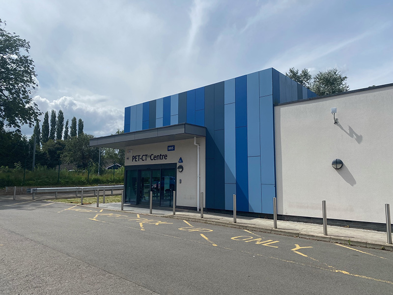
Leicester PET/CT Centre
The NHS England contract to supply PET/CT services is provided by Alliance Medical Ltd (AML). Alliance Medical Ltd have installed a static PET/CT Unit at the Glenfield Hospital site. Scientific support is provided by the UHL Nuclear Medicine Service and appointment bookings are undertaken by Alliance Medical Ltd.
A PET/CT scan is a medical imaging technique used to produce a detailed, three-dimensional picture of the inside of the body. A PET/CT scan involves injecting a small amount of a radioactive tracer into a vein, usually in your arm. The most commonly used tracer in PET/CT is FDG (fluoro-deoxy-glucose),which is similar to naturally occurring glucose (a type of sugar) so your body treats it in a similar way.
The scanner detects the radioactive tracer as it collects in different parts of your body. By analysing the areas where the radiotracer does and does not build up, it’s possible to shows how the body breaks down and uses glucose. Abnormal cells use glucose differently and this will show up on the scan.
The radioactive tracers used for your scan have a short shelf life and has to be made on the day of your scan. Therefore occasionally appointments may be cancelled at short notice if there are tracer production issues. A typical PET/CT scan takes approximately 2 hours.
Do NOT have anything to eat for six hours before your appointment. During this time you can ONLY drink unflavoured water. For your scan, you do not need to have a full bladder. You should avoid strenuous exercise for 24 hours before your appointment. If you smoke or vape, withhold from smoking or vaping 2hours before your appointment.
It’s a good idea to wear loose, comfortable clothes. Avoid wearing jewelry and clothes that have metal parts, such as zips, because these will need to be removed.
When you arrive at the department, we will ask you to complete safety questionnaire.
You will when be taken to a preparation room to sit or lie on a couch and a small cannula will be inserted into a vein in your arm. Through the cannula we will then administer a small dose of a radioactive tracer and ask you to remain lying down for about one hour before your scan.
After one hour, we will ask you to empty your bladder before taking you through to the scanning room and onto the scanning bed.
The scan can take between 25 to 45 minutes depending on the type of scan your doctor has requested.
You will be asked to have either your arms above your head or down by your side.
It is important that you remain still during your scan.
Once the scan is completed, you will be able to leave the department immediately.
You will be able to eat and drink as you normally do. You may go anywhere you wish but you should avoid close contact with children and pregnant women for 8 hrs after your scan. This is to avoid exposing children to unnecessary radiation.
Your PET/CT scan will be reported within 3-5 working days. The results of your scan will then be made available to the referring doctor.
We will expose you to small level of ionising radiation when we carry out this examination.
We are all exposed to ionising radiation from naturally occurring sources such as cosmic rays, certain types of soil and rocks and even food we eat. Ionising radiation can cause cell damage that in turn, after many years, may turn cancerous.
The radiation associated with your exam will therefore carry a small risk which is less than 0.1%. This risk will be far outweighed by the benefits of having this exposure. We will also tailor the amount of radiation we use for you.
Phone number: 0116 258 3220
Email Address: [email protected]
Address
PET CT Centre (opposite South Entrance of Glenfield Hospital)
Glenfield Hospital
Groby Road
Leicester
LE3 9QP
Opening times
Monday: 7:30am to 7:30pm
Tuesday: 7:30am to 7:30pm
Wednesday: 7:30am to 7:30pm
Thursday: 7:30am to 7:30pm
Friday: 7:30am to 7:30pm
We provide several easy options for submitting Nuclear Medicine referrals. Please choose the method that works best for you:
- Submit via ICE or NerveCentre
- Or complete a Nuclear Medicine Request Form below and email to:
Inpatient Referrals
- Please use ICE or NerveCentre wherever possible to avoid delays
- Alternatively, complete a Nuclear Medicine Request Form below and email to:
PET-CT Requests
- Submit via ICE or NerveCentre
- Or complete a Nuclear Medicine Request Form and email to:
Download link for:
MPNMNM0268 – Nuclear Medicine Request Form v2.0 (271kB pdf)
Blank PET Referral Form 2015-04 (193kB pdf)
MPNMNM0285 – BSLNB request form GGH v1.2 (297kB pdf)
Need help? Contact our team
Leicester Royal Infirmary: 0116 258 5627
Glenfield Hospital: 0116 258 3850